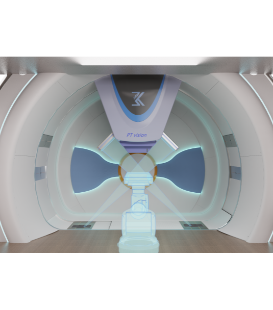


In proton therapy, precise positioning is essential to ensure accurate and effective treatment. The PT vision CBCT Image-Guided System uses cone-beam X-ray projection to scan the patient and reconstruct high-resolution, 3D volumetric images through advanced computer processing. PT vision enables accurate visualization of anatomical structures and is widely applicable in medical scenarios such as cranial, thoracic, and abdominal imaging, as well as radiation therapy planning.


| Effective Imaging Area | 430x430 mm2 |
| CBCT Imaging Field of View | Full-fan:φ310*300 Half-fan:φ550*245 |
| Image Registration | Manual and automatic |
| Auto Registration Time | ≤10 s |
| Registration Correction Vector | Supports 6 degrees of freedom (6DOF) |
| Positioning Accuracy | ≤1 mm |
| Source-to-Axis Distance (SAD) | 2800±0.5 mm |
| Source-to-Image Distance (SID) | 3800±0.5 mm |
| Reconstruction Time | Full-fan (200°):≤45 s Half-fan (360°):≤75 s |
| Imaging Dose (at 100 kV) | 65.04 mGy / 100 mAs / 400 frames |

Address:No.2299 Boyanwan Road, Hefei Hi-Tech Development Zone, Hefei, Anhui, China

Follow us

 Products &Solution